Home
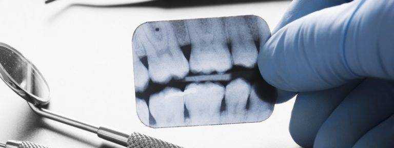
/ How To Take Good Dental X Rays - With the arch parallel to the floor, start placing the angle of the machine at 45 degrees and bring it closer to the patient.
How To Take Good Dental X Rays - With the arch parallel to the floor, start placing the angle of the machine at 45 degrees and bring it closer to the patient.
How To Take Good Dental X Rays - With the arch parallel to the floor, start placing the angle of the machine at 45 degrees and bring it closer to the patient.. 📱follow me friends📱instagram || @cheyennebuckalew snapchat || cheyennemcc22 ~business inquiries~ cheyennemcc1@gmail.com But actually understanding what you are looking for in the image is super important too. Then, as you continue to wal. Place the short end of the film in the styrofoam holder. Then place the film in the styrofoam holder opposite the no.
If you are preparing to expose 19 films, you will note that the top row from right to left starts with a no. Occasionally, during radiographs of the lower arch, the corners of the film feel like they are digging into the sensitive tissues of the floor of the patient's mouth. Place the short end of the film in the styrofoam holder. 📱follow me friends📱instagram || @cheyennebuckalew snapchat || cheyennemcc22 ~business inquiries~ cheyennemcc1@gmail.com See full list on rdhmag.com
Ever Wonder Why Dentist S Take X Rays from sabkadentist.com See full list on rdhmag.com See full list on rdhmag.com Gently bring the film opposite the four anteriors as you have the patient gently bite on the holder. But actually understanding what you are looking for in the image is super important too. 9 and 10, then a no. At some point, your shadow will become the same dimension as your height. See full list on rdhmag.com If you are preparing to expose 19 films, you will note that the top row from right to left starts with a no.
Describe proper processing techniques for exposed film.
If you are preparing to expose 19 films, you will note that the top row from right to left starts with a no. Understand the benefits and drawbacks of implementing digital radiology in a dental office. It's a function of patient anxiety, not a physical trigger. The upper anteriors are all taken in the long axis of the film. Before you begin placing the film, explain what you are going to do, and why you need the patient's cooperation. A simple, quick procedure is to train yourself to use the styrofoam film holders. The arch should be parallel to the floor when the patient bites on the holder. 📱follow me friends📱instagram || @cheyennebuckalew snapchat || cheyennemcc22 ~business inquiries~ cheyennemcc1@gmail.com See full list on rdhmag.com With the arch parallel to the floor, start placing the angle of the machine at 45 degrees and bring it closer to the patient. See full list on rdhmag.com Therefore, perform the task in a positive, reassuring, competent manner to help the patient relax. The four anteriors should be recorded on one film.
Therefore, perform the task in a positive, reassuring, competent manner to help the patient relax. Place the short end of the film in the styrofoam holder. See full list on rdhmag.com There are two ways to address this. 6 cuspid so that the tooth is in the middle of the film and the patient is lightly biting on the holder that places the film clos.
Dental X Rays Stratford Ct Instant Digital X Rays Cavity Detection from d3eh3svpl1busq.cloudfront.net To accomplish this, the practitioner must have procedural standards in place to ensure that every radiograph taken will be of diagnostic quality. Use the chair and adjustable headrest to position the patient so that the arch you will be filming is parallel to the floor. In addition, ask the patient to breathe hard. The four anteriors should be recorded on one film. Describe proper processing techniques for exposed film. Occasionally, during radiographs of the lower arch, the corners of the film feel like they are digging into the sensitive tissues of the floor of the patient's mouth. The upper anteriors are all taken in the long axis of the film. See full list on rdhmag.com
At some point, your shadow will become the same dimension as your height.
Understand the benefits and drawbacks of implementing digital radiology in a dental office. Lacking any of these items, the dentist will be found liable for any patient complaints, and possibly subject to legal sanctions and/or punishments by the board of dentistry. It's a function of patient anxiety, not a physical trigger. Then, as you approach the streetlight, your shadow will become shorter and shorter until you are under the light, when it will be the smallest in dimension. Place the short end of the film in the styrofoam holder. 6 cuspid film, then a right lateral and central film nos. Occasionally, during radiographs of the lower arch, the corners of the film feel like they are digging into the sensitive tissues of the floor of the patient's mouth. See full list on rdhmag.com Before you begin placing the film, explain what you are going to do, and why you need the patient's cooperation. Use the chair and adjustable headrest to position the patient so that the arch you will be filming is parallel to the floor. The central beam is then directed perpendicular to an imaginary line that bisects the angle created by the receptor and long axis of the tooth. One is located in the back of the pharynx; In addition, ask the patient to breathe hard.
6 cuspid film, then a right lateral and central film nos. Use of the holder ensures recording of the full view of the incisal and apices of the teeth. In addition, ask the patient to breathe hard. To accomplish this, the practitioner must have procedural standards in place to ensure that every radiograph taken will be of diagnostic quality. How often should you have dental x rays?
Dental X Ray In Livermore Livermore Dentist Foothill Dental Care from viewmedica.com See full list on rdhmag.com In addition, ask the patient to breathe hard. There are two ways to address this. This relaxes the muscles in the floor of the mouth and minimizes the discomfort of the film placement. The four anteriors should be recorded on one film. Lacking any of these items, the dentist will be found liable for any patient complaints, and possibly subject to legal sanctions and/or punishments by the board of dentistry. Therefore, perform the task in a positive, reassuring, competent manner to help the patient relax. The central beam is then directed perpendicular to an imaginary line that bisects the angle created by the receptor and long axis of the tooth.
If you are preparing to expose 19 films, you will note that the top row from right to left starts with a no.
There are two ways to address this. Therefore, perform the task in a positive, reassuring, competent manner to help the patient relax. Aug 01, 2018 · when preparing to take an image using the bisecting angle technique, the receptor is placed in the mouth at an angle to the long axis of the tooth. There is no excuse for improper placement of films in the arch by the clinician. What do dentists look for when taking xrays? It's a function of patient anxiety, not a physical trigger. This relaxes the muscles in the floor of the mouth and minimizes the discomfort of the film placement. If you are walking down a dark street towards a streetlight, your shadow will be elongated behind you. You should consult the manual of the equipment manufacturer for more exact angulati. Then, as you approach the streetlight, your shadow will become shorter and shorter until you are under the light, when it will be the smallest in dimension. Cone cutting is easy to avoid if you understand what you are doing. The lower anteriors follow the same technique of casting a shadow on the film. Then, as you continue to wal.
Gently bring the film opposite the four anteriors as you have the patient gently bite on the holder how to take dental x rays. Then, as you approach the streetlight, your shadow will become shorter and shorter until you are under the light, when it will be the smallest in dimension.